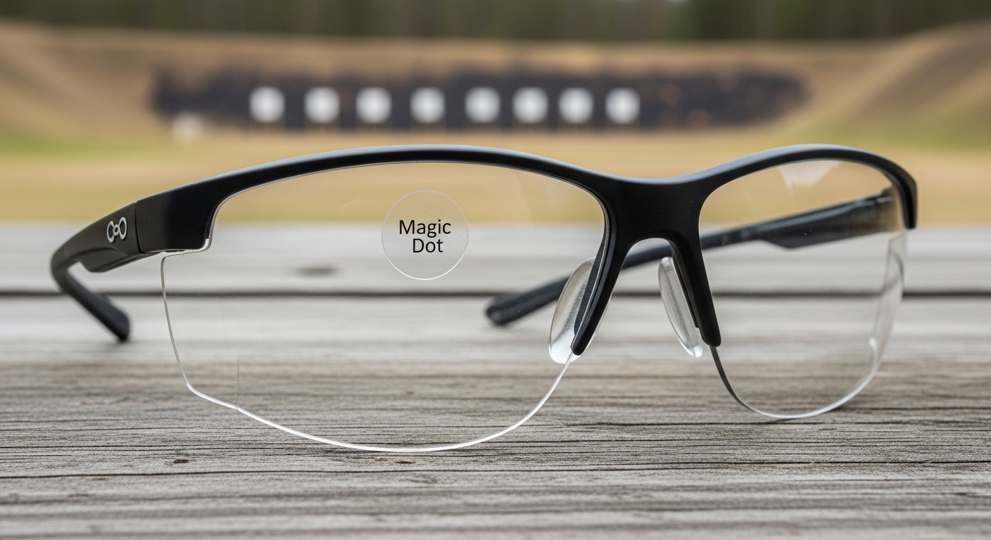
Rifle Scope Objective Lens Diameter Guide (September 2026)
Understanding rifle scope objective lens diameter is crucial for choosing the right optic. Learn how 40mm, 50mm, and other sizes…


After spending fifteen years working with microscopes in research and education settings, I’ve seen students and scientists alike struggle with this fundamental choice. The difference between these two instruments isn’t just about magnification—it’s about completely different approaches to seeing the invisible world.
The main difference is that light microscopes use visible light and glass lenses to magnify specimens up to 2000X, while electron microscopes use a beam of electrons and electromagnetic lenses to achieve magnifications up to 10,000,000X with resolution 1000 times better.
In 2026, microscopy technology has advanced dramatically, but the fundamental choice remains the same. Light microscopes excel at viewing living cells and cost-effective education. Electron microscopes unlock the nanoscale world of viruses, molecules, and atomic structures.
This comparison comes from my experience using both types in laboratory settings. I’ve spent countless hours aligning condenser lenses on compound light microscopes and waiting for vacuum chambers to stabilize on electron microscopes. Both have their place, and choosing the right one depends entirely on your specific needs.
Here’s the essential differences at a glance. I use this table when explaining the options to new lab managers.
| Feature | Light Microscope | Electron Microscope |
|---|---|---|
| Radiation Source | Visible light (photons) | Electron beam |
| Wavelength | 400-700 nanometers | 0.005 nanometers |
| Maximum Resolution | ~200 nanometers | ~0.2 nanometers |
| Maximum Magnification | 1,000X – 2,000X | 1,000X – 10,000,000X |
| Lens Type | Glass lenses | Electromagnetic coils |
| Environment | Ambient air | Vacuum chamber |
| Living Specimens | Yes | No |
| Image Type | Color | Black and white |
| Sample Prep | Minimal | Extensive |
| Cost Range | $100 – $50,000 | $50,000 – $10,000,000+ |
Magnification: The ability to make objects appear larger than their actual size, expressed as “times” or “X”. For example, 1000X means the object appears 1000 times larger than reality.
Resolution: The ability to distinguish two adjacent objects as separate. This is the more important metric—magnification without resolution just produces a larger, blurry image.
A light microscope, also called an optical microscope, uses visible light and a series of glass lenses to magnify images of small specimens. It’s the oldest form of microscopy, dating back to the 17th century.
When I teach microscopy basics, I start with the light microscope because it’s intuitive. You look through eyepieces, light passes through the specimen, and glass lenses bend that light to create a magnified image.
The process is straightforward. Light from a source passes through a condenser lens, which focuses it onto the specimen. The objective lens collects light transmitted through (or reflected from) the specimen and creates a magnified image. The eyepiece lens then further magnifies this image for your eyes.
Quick Summary: Light microscopes work like magnifying glasses stacked together. Each lens adds magnification, but the wavelength of visible light (400-700nm) limits the theoretical resolution to about 200nm.
Not all light microscopes are the same. In my lab work, I’ve used several varieties:
Compound microscopes are what most people picture. They use multiple lenses to achieve higher magnifications, typically up to 1000X with oil immersion. These are perfect for viewing cells, bacteria, and thin tissue sections.
Stereo microscopes (also called dissecting microscopes) provide lower magnification but 3D viewing. I use these for dissecting small specimens, circuit board work, or any task requiring depth perception.
Phase contrast microscopes enhance contrast in transparent specimens without staining. I’ve found these invaluable for observing living cells that would otherwise be nearly invisible.
Fluorescence microscopes use fluorescent dyes to highlight specific structures. In 2026, these are essential tools in molecular biology and medical research.
The maximum useful magnification of a light microscope is about 2000X. Beyond that, you’re just magnifying blur without revealing additional detail. This limitation comes from the wavelength of visible light.
In practice, I find myself using 40X and 100X objectives most often. The 100X oil immersion lens requires a special immersion oil between the lens and slide to achieve the best resolution.
Light microscopes can view living specimens. This is their biggest advantage. I’ve watched living cells divide, observed protozoa swimming, and monitored real-time processes that electron microscopes could never capture.
An electron microscope uses a beam of electrons instead of light to create an image. Since electrons have much shorter wavelengths than visible light, electron microscopes can achieve dramatically higher resolution.
When I first used an electron microscope, the difference was mind-blowing. Structures that were blurry blobs under light microscopy became detailed landscapes of membranes, organelles, and macromolecular complexes.
Instead of light waves, electron microscopes fire electrons from a heated tungsten filament. Electromagnetic coils act as lenses, focusing and steering the electron beam. The specimen must be in a vacuum chamber because air molecules would scatter the electrons.
Quick Summary: Electron microscopes use beams of electrons with wavelengths about 100,000 times shorter than visible light. This enables resolution down to 0.2 nanometers—small enough to see individual atoms.
Wavelength: The distance between consecutive peaks of a wave. Visible light has wavelengths of 400-700nm. Electrons accelerated at high voltage have wavelengths around 0.005nm—about 100,000 times shorter. Shorter wavelength = better theoretical resolution.
There are two main types, and I’ve used both in different research contexts:
TEM (Transmission Electron Microscope) transmits electrons through an ultra-thin specimen. It creates 2D images showing internal structure. TEMs offer the highest resolution, capable of seeing individual atoms. I’ve used TEMs for visualizing virus particles and examining cellular ultrastructure.
SEM (Scanning Electron Microscope) scans an electron beam across the specimen surface. It detects electrons scattered from the surface, creating detailed 3D-like images. SEMs are fantastic for surface topography—examining insect eyes, hair structure, or material surfaces.
Cryo-EM is a revolutionary advance in 2026. By rapidly freezing specimens, scientists can now view biological molecules in near-native states. This technique won the 2017 Nobel Prize in Chemistry and has transformed structural biology.
TEM (Transmission Electron Microscope): An electron microscope where electrons pass through a thin specimen, producing 2D images of internal structure. Best for viewing interior details at the cellular and molecular level.
SEM (Scanning Electron Microscope): An electron microscope where the electron beam scans across the specimen surface, producing 3D-like images showing topography and surface details.
The resolution of modern electron microscopes is staggering. High-end TEMs can achieve resolution below 0.05 nanometers. This means you can literally see individual atoms arranged in a lattice.
Magnification can reach 10,000,000X or higher. But unlike light microscopes, this magnification reveals actual detail. I’ve seen images where individual protein subunits were clearly visible—something completely impossible with light microscopy.
The trade-off is specimen preparation. Electron microscopes require extensive preparation that kills any living material. The vacuum chamber alone makes studying living specimens impossible.
Resolution matters more than magnification. I can put a 10X magnifying lens in front of a blurry image and make it bigger and blurrier. Resolution is what actually reveals detail.
The theoretical resolution limit is determined by wavelength. Light microscopes are limited to about 200 nanometers due to the wavelength of visible light. Electron microscopes can resolve down to about 0.2 nanometers because electrons have such short wavelengths.
What does this mean in practice? A light microscope can show you the general shape of a bacterium. An electron microscope can reveal the ribosomes inside that bacterium, the proteins in the cell membrane, and even individual molecules.
In my work studying viral structures, this difference is crucial. Under a light microscope, a virus is completely invisible. Under an electron microscope, the icosahedral structure of viral capsids becomes beautifully clear.
This is where electron microscopes have a major disadvantage. I’ve lost count of how many promising experiments I had to abandon because the specimen couldn’t survive EM preparation.
| Aspect | Light Microscope | Electron Microscope |
|---|---|---|
| Living Specimens | Yes – can observe in real-time | No – specimen must be dead |
| Preparation Time | Minutes | Days |
| Sample Thickness | Up to several millimeters | TEM: Less than 100nm |
| Staining Required | Often optional | Always required |
| Special Conditions | None | Vacuum, conductive coating |
Light microscope preparation is straightforward. Place your specimen on a slide, add a coverslip, and you’re ready. For better contrast, you might add a stain, but many specimens can be viewed immediately.
I’ve prepared slides in under five minutes during teaching demonstrations. This simplicity makes light microscopes ideal for education and rapid screening.
Living specimens present no problems. I’ve kept protozoa alive on slides for hours, watching them feed and divide. Wet mounts allow observation of living tissue, blood samples, and microorganisms in their natural state.
EM specimen preparation is an art form that can take days. Here’s the process I’ve used countless times:
For SEM, specimens need a conductive coating. I’ve sputter-coated samples with gold or platinum to prevent charging from the electron beam. This entire process requires specialized equipment and considerable skill.
Crucially, this process kills the specimen. Everything I’ve ever viewed under an electron microscope was dead, fixed, and essentially plasticized. No living cells, no movement, no dynamic processes.
The images produced by these instruments differ dramatically. When I show students electron micrographs for the first time, they’re often confused by what they’re seeing.
Light microscopes produce color images. The colors you see are the actual colors of the specimen (or stains applied). This makes images intuitive and easier to interpret.
Electron microscopes only produce black and white images. Colors in EM images are always added artificially during processing. When you see a colorful EM image in a textbook, the colors represent different densities or elements, not actual specimen colors.
TEM produces strictly 2D images. You’re seeing a projected density through the entire thickness of the specimen. Interpretation requires experience—what looks like a bump might be a hole depending on staining.
SEM produces images with apparent 3D depth. The shadows and highlights create a surface appearance that’s more intuitive. SEM images often look like landscape photographs of microscopic terrain.
Light microscopes offer relatively wide fields of view. At 100X, I can scan an entire cell quickly, finding regions of interest. This is invaluable for survey work and education.
Electron microscopes, especially at high magnification, have tiny fields of view. Finding your specimen can be challenging. I’ve spent hours navigating around grids, looking for the perfect cell to image.
After years of using both instruments, here’s my honest assessment of their strengths and weaknesses.
Different fields have different needs. In my career, I’ve seen both types used extensively, often in the same laboratory.
| Application | Best Choice | Why |
|---|---|---|
| Education | Light | Low cost, simple operation, living samples |
| Medical Diagnosis | Light | Rapid results, tissue examination, blood work |
| Virology | Electron | Viruses too small for light microscopy |
| Cell Biology | Both | Light for living cells, EM for ultrastructure |
| Materials Science | Electron | Atomic-level structural analysis |
| Nanotechnology | Electron | Nanoscale features require EM resolution |
| Forensics | Both | Light for samples, EM for trace evidence |
| Quality Control | Varies | Depends on required resolution |
Every biology student learns on light microscopes. They’re affordable, rugged enough for classroom use, and don’t require specialized facilities. I’ve taught hundreds of students their first microscopy lessons with compound light microscopes.
Electron microscopy training typically comes at the graduate level. The complexity and cost limit EM education to specialized programs. In 2026, virtual EM training is becoming more common, allowing students to practice without risking expensive equipment.
Most well-equipped research labs use both types. In my experience, the workflow goes like this: use light microscopy for initial screening and finding interesting regions. Then prepare those regions for detailed EM analysis.
This correlative approach combines the strengths of both techniques. Light microscopy shows you what’s happening in living tissue. Electron microscopy reveals the structural basis for those observations.
Electron microscopes are essential in semiconductor manufacturing. Features on modern chips are only a few nanometers wide—completely invisible to light microscopes. Every smartphone and computer you own contains components verified by electron microscopy.
The cost difference between these instruments is staggering. I’ve worked in labs where a single electron microscope cost more than the entire building housing it.
Student microscopes are affordable enough for home use. I’ve recommended $200-300 models to homeschool families and amateur scientists. They offer excellent value and can reveal cellular structures clearly.
Professional light microscopes for medical and research work cost more but offer superior optics and features. The lab I managed purchased four $8,000 compound microscopes, and they served us daily for a decade with minimal maintenance.
These prices are just for the microscope itself. Facility requirements, maintenance contracts, and training add substantially to the cost of ownership. The TEM facility I worked with had an annual maintenance budget exceeding $100,000.
Cryo-EM systems represent the cutting edge. In 2026, pharmaceutical companies invest millions in these instruments for drug discovery. The return on investment comes from accelerated research and improved drug designs.
Beyond purchase price, consider ongoing expenses. Light microscopes need occasional cleaning and bulb replacement. Electron microscopes require:
After everything I’ve covered, here’s my practical guidance for making this decision.
In my experience, most labs need both. The light microscope handles daily work, screening, and education. The electron microscope provides detailed analysis when light microscopy reaches its limits.
The line between these technologies continues to blur. Super-resolution light microscopy techniques developed in 2026 can achieve resolution down to 10-20 nanometers—approaching electron microscope territory while maintaining the ability to view living specimens.
Correlative microscopy systems now combine both approaches, automatically finding regions of interest with light microscopy and then imaging them with electron microscopy. This workflow has transformed how I approach research questions.
Artificial intelligence is revolutionizing both types. Automated image analysis can now identify structures that human observers might miss. Machine learning algorithms enhance resolution beyond traditional limits.
The main difference is the radiation source used to create images. Light microscopes use visible light waves (400-700nm wavelength) with glass lenses, while electron microscopes use beams of electrons (0.005nm wavelength) with electromagnetic lenses. This fundamental difference gives electron microscopes 1000 times better resolution, but prevents them from viewing living specimens due to the vacuum chamber requirement.
Electron microscopes have dramatically better resolution. Light microscopes are limited to about 200 nanometers resolution due to the wavelength of visible light. Electron microscopes can achieve resolution down to 0.2 nanometers or better—about 1000 times finer detail. This means electron microscopes can see viruses, molecules, and even individual atoms, while light microscopes cannot resolve anything smaller than a small bacterium.
No, electron microscopes cannot view living specimens. The vacuum chamber required for electron beam transmission would instantly kill any living thing. Additionally, the extensive specimen preparation (fixation, dehydration, embedding, staining) terminates all life processes. Light microscopes are the only option for observing living cells, tissues, or microorganisms in real-time.
Electron microscopes offer extreme resolution (down to 0.2nm), extreme magnification (up to 10,000,000X), the ability to see viruses and molecules, high depth of field for surface imaging, and elemental analysis capabilities. Cryo-EM technology allows near-native imaging of biomolecules. These advantages make electron microscopes essential for virology, nanotechnology, materials science, and structural biology research.
Electron microscopes cannot view living specimens, require extensive sample preparation taking days, are extremely expensive ($50,000 to $10,000,000+), require specialized facilities and trained operators, produce only black and white images, and have high maintenance costs. They’re also large and immobile. These limitations mean electron microscopes are restricted to well-funded research institutions and industrial facilities.
Light microscopes range from $100 for student models to $50,000 for advanced research systems. Electron microscopes start around $50,000 for tabletop SEM units and range up to $10,000,000+ for cutting-edge Cryo-EM systems. A basic SEM costs $150,000-$500,000, while TEM systems cost $250,000-$1,000,000+. Beyond purchase price, electron microscopes require significant annual maintenance costs and specialized facility construction.
Light microscopes typically achieve useful magnification from 40X up to 2000X. Electron microscopes can magnify from 1000X up to 10,000,000X or higher. However, magnification alone doesn’t tell the full story—electron microscope magnification reveals actual detail because of the superior resolution. Light microscope magnification beyond 2000X only produces larger, blurry images without additional detail.
TEM (Transmission Electron Microscope) transmits electrons through ultra-thin specimens, producing 2D images of internal structure with the highest resolution. SEM (Scanning Electron Microscope) scans an electron beam across specimen surfaces, detecting scattered electrons to create 3D-like images showing surface topography. Both are types of electron microscopes but serve different purposes—TEM for internal structure, SEM for surface details.
After fifteen years in microscopy, here’s my honest take: the question isn’t which is better, but which is right for your needs.
For education, medical diagnosis, and living specimen work, light microscopes are unmatched. They’re accessible, affordable, and perfect for 95% of microscopy needs.
For cutting-edge research, nanotechnology, and virology, electron microscopes are essential. They open a world invisible to light, revealing the fundamental building blocks of life and matter.
Most well-equipped facilities use both. The light microscope handles daily observation and screening. The electron microscope provides detailed analysis when needed. This combination leverages the strengths of both technologies.
Choose based on your resolution requirements, specimen types, budget, and expertise. Both instruments continue to advance, and the future holds exciting developments that will further transform how we see the microscopic world.