Night Vision 101: Complete Beginner’s Guide 2026
Complete beginner's guide to night vision technology. Learn how image intensification works, understand specifications like FOM and SNR, compare generations,…


I’ve helped hundreds of students and hobbyists choose their first microscope over the past 15 years. The most common mistake I see? Someone buying a compound microscope when they really needed a stereo microscope (or vice versa). This confusion costs $100-300 per mistake, and the wrong microscope ends up gathering dust in a closet.
Let me clear up the confusion immediately.
The main difference: A compound microscope uses high magnification (40x-1000x) to view thin, transparent specimens on slides with light from below, while a stereo microscope uses lower magnification (10x-40x) to view 3D objects with light from above. Compound microscopes are for cells and bacteria. Stereo microscopes are for coins, insects, and electronics.
This distinction matters because choosing the wrong tool means you literally cannot see what you want to study. A stereo microscope cannot show you bacteria. A compound microscope cannot show you the details on a coin in 3D.
Compound microscopes are for seeing tiny things (cells, bacteria) on flat slides. Stereo microscopes are for seeing larger 3D things (coins, insects, electronics) with depth perception.
| Feature | Compound Microscope | Stereo Microscope |
|---|---|---|
| Magnification Range | 40x to 1000x+ | 10x to 40x (some up to 80x) |
| View Type | 2D flat image | 3D stereo image |
| Light Source | From below (transmitted) | From above (reflected) |
| Specimen Type | Thin, transparent on slides | Opaque 3D objects |
| Working Distance | Very short (mm range) | Long (room to work) |
| Depth of Field | Shallow | Deep |
| Best For | Cells, bacteria, pond water | Dissection, coins, electronics |
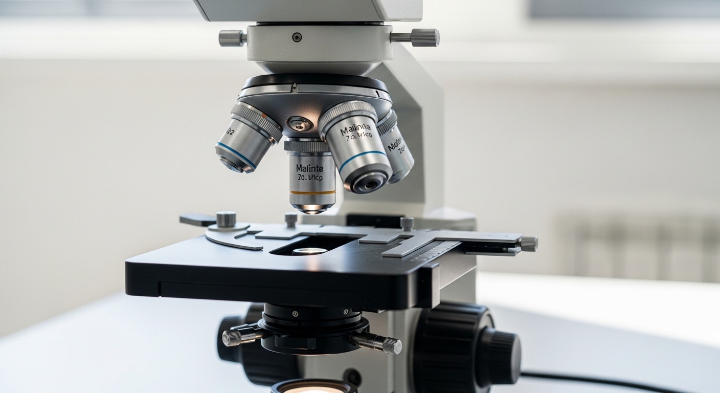
A compound microscope is what most people picture when they hear “microscope.” It’s the classic microscope with multiple objective lenses on a rotating nosepiece. I’ve used these extensively in biology labs and seen thousands of students learn with them.
Compound Microscope: An optical microscope that uses two sets of lenses (objective and eyepiece) to achieve high magnification. Light passes through thin specimens from below, making them ideal for transparent samples.
Compound microscopes use transmitted light. The light source is built into the base, shining upward through the stage. Your specimen sits on a glass slide, and light passes through it into the objective lenses. This optical system is why specimens must be thin and transparent.
The magnification system works by multiplying two lens powers. If you use a 10x eyepiece with a 40x objective, you get 400x total magnification. Most compound microscopes come with three or four objective lenses: typically 4x, 10x, 40x, and 100x (the 100x requires oil immersion).
| Objective Lens | Eyepiece | Total Magnification | Best For |
|---|---|---|---|
| 4x (scanning) | 10x | 40x | Finding specimens, overview |
| 10x (low power) | 10x | 100x | General observation |
| 40x (high dry) | 10x | 400x | Cell details, nuclei |
| 100x (oil immersion) | 10x | 1000x | Bacteria, fine detail |
Compound microscopes require prepared specimens. You cannot just drop an insect under a compound microscope and expect to see anything useful. The specimen must be:
I’ve spent many hours preparing slides in the lab. A drop of pond water on a slide with a coverslip is the simplest preparation. More complex specimens require fixing, sectioning with a microtome, and staining with specific dyes.
A stereo microscope (also called a dissecting microscope) provides a completely different viewing experience. Instead of looking at a flat, magnified image, you see a three-dimensional view with depth perception. I’ve used stereo microscopes for everything from dissection work to examining camera sensor components.
Stereo Microscope: Also known as a dissecting microscope, it uses two separate optical paths (one for each eye) to create a 3D image. Light reflects off the specimen from above, making it ideal for opaque objects.
Stereo microscopes use reflected light. The light source (or lights) shines down onto the specimen from above. Light bounces off the object and enters the two separate optical paths, creating the stereoscopic (3D) effect that gives this microscope its name.
Because you’re not looking through a specimen, magnification is much lower. But here’s the key advantage: you get a long working distance. This means there’s actual space between the objective lens and your specimen. You can manipulate your sample while viewing it.
The stereo effect comes from having two slightly different angles of view, just like your eyes perceive depth in the real world. Each eyepiece receives its own image through a separate optical path.
This 3D capability makes stereo microscopes incredibly practical. When you’re dissecting a frog or soldering a small component, you need depth perception. You need to know how far your tools are from the specimen. A compound microscope cannot provide this.
Stereo microscopes typically offer 10x to 40x magnification. Some models go up to 80x, but beyond that the stereo effect becomes less effective and the shallow depth of field negates the advantages.
| Magnification | Working Distance | Best Applications |
|---|---|---|
| 10x | 75-100mm | Large dissection, sorting |
| 20x | 50-75mm | General inspection, coin details |
| 30x | 35-50mm | Electronics, small components |
| 40x | 25-35mm | Fine detail work |
This is the huge advantage of stereo microscopes. Just place your object on the stage and start viewing. No slides, no coverslips, no staining, no sectioning. This makes stereo microscopes much more beginner-friendly and practical for many applications.
Understanding the technical differences helps solidify which microscope suits your needs. Let me break down the most important distinctions.
Higher magnification is not always better. It depends on what you’re trying to see. Compound microscopes offer much higher magnification but lose the 3D perspective. Stereo microscopes provide lower magnification but with realistic depth perception.
I’ve seen students disappointed when they put a whole insect under a compound microscope at 400x. They see nothing but black because light cannot penetrate the specimen. The same insect under a stereo microscope at 20x reveals amazing details.
Working distance is the space between the objective lens and your specimen. This practical consideration matters for real work.
Working Distance: The distance between the front of the microscope objective lens and the focal point on the specimen. Longer working distance allows you to manipulate the specimen while viewing.
Compound microscopes have extremely short working distances, often just a few millimeters. You cannot work on your specimen while viewing. You look, then you remove the slide to work.
Stereo microscopes have generous working distances. At 20x magnification, you might have 60mm of clearance. You can dissect, solder, or manipulate while continuously viewing. This is crucial for many practical applications.
Depth of field refers to how much of your specimen is in focus at once. Higher magnification means shallower depth of field. This is a fundamental trade-off in optics.
With a compound microscope at 1000x, only a thin slice of your specimen is in focus at any time. You must constantly adjust the focus knob to see different planes.
Stereo microscopes at 20x have much greater depth of field. More of your 3D specimen remains in focus simultaneously, making the viewing experience more natural and useful.
The lighting direction is fundamentally different and relates to specimen type:
Some advanced stereo microscopes offer both transmitted and reflected illumination. This dual lighting is useful for translucent specimens like thin plant sections where you want both surface detail and some internal visibility.
Generally speaking, stereo microscopes cost more than compound microscopes at comparable quality levels. The dual optical path and precision mechanics required for stereo viewing add to the manufacturing cost.
Entry-level compound microscopes start around $80-150. Entry-level stereo microscopes typically begin around $150-200. Professional models of either type can cost thousands of dollars.
Let me make this decision simple based on what you want to do.
If you’re buying a first microscope for a child or student, consider their interests. A student interested in nature and biology will likely prefer a compound microscope. A student interested in tinkering, electronics, or collecting will prefer a stereo microscope.
Elementary students often find stereo microscopes more engaging. They can just place objects under the microscope and see interesting details immediately. No slide preparation means faster gratification.
For middle school and above, compound microscopes align better with science curricula and offer more educational value for biology studies.
Many professionals end up owning both. I’ve known entomologists who use stereo microscopes for specimen identification and compound microscopes for examining insect structures at the cellular level.
Your specific application should drive the decision. Don’t let higher magnification numbers sway you toward a compound microscope if you primarily work with 3D objects.
The main difference is magnification and specimen type. Compound microscopes provide high magnification (40x-1000x) for viewing thin, transparent specimens on slides with transmitted light. Stereo microscopes provide lower magnification (10x-40x) for viewing 3D opaque objects with reflected light, giving you a three-dimensional view.
A compound microscope is best for viewing cells. Cells are thin and semi-transparent, allowing light to pass through them. Compound microscopes provide the necessary magnification (400x-1000x) to see cell structures like nuclei, organelles, and cell walls clearly.
No, you cannot see bacteria with a stereo microscope. Bacteria are too small and require magnification of at least 400x, typically 1000x with oil immersion. Stereo microscopes only magnify 10x-40x, which is insufficient for viewing bacteria.
Stereo microscopes are used for examining 3D objects like insects, coins, jewelry, electronics, and plants. They’re essential for dissection work, electronics repair and soldering, quality control inspection, forensics, and any application where you need to manipulate the specimen while viewing.
With a compound microscope you can see plant and animal cells, bacteria, protozoa, blood cells, pond water organisms, fungi, thin tissue sections, and prepared slides. You’ll need magnification of 400x-1000x to clearly see cell structures and bacteria details.
Use a compound microscope when viewing thin, transparent specimens on slides. This includes cells, bacteria, pond water samples, blood smears, and prepared tissue sections. Choose a compound microscope for biology education, medical diagnosis, microbiology research, and any application requiring high magnification of microscopic structures.
Use a stereo microscope when examining whole 3D objects. This includes dissection work, electronics inspection and soldering, coin and stamp collecting, jewelry examination, insect study, plant inspection, and any application where you need to manipulate your specimen while viewing or want depth perception.
No, stereo microscopes have lower magnification than compound microscopes. Stereo microscopes typically provide 10x-40x magnification, while compound microscopes provide 40x-1000x or more. However, stereo microscopes provide 3D viewing and longer working distance, which compound microscopes cannot offer.