How to Read Mirage With a Spotting Scope
Learn to read mirage with a spotting scope for accurate wind calls. Complete guide covering equipment setup, step-by-step techniques, mirage…

Choosing between an inverted and upright microscope can make or break your research workflow. After working with both types in various lab settings, I’ve seen labs waste thousands of dollars on the wrong configuration.
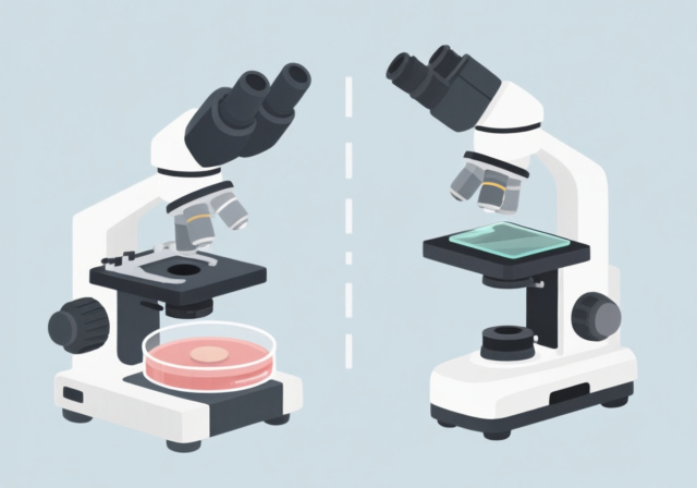
The main difference lies in objective lens placement: inverted microscopes have objectives below the stage pointing upward, while upright microscopes have objectives above the stage pointing downward. Choose inverted for live cell cultures in dishes and upright for fixed specimens on glass slides.
This fundamental difference in design dictates everything from sample preparation to long-term maintenance costs. Let me break down exactly what you need to know.
| Feature | Inverted Microscope | Upright Microscope |
|---|---|---|
| Objective Position | Below stage, pointing upward | Above stage, pointing downward |
| Condenser Location | Above the stage | Below the stage |
| Sample Placement | On top of stage (dishes, flasks, plates) | Between stage and objective (slides) |
| Best Sample Types | Live cells, cell cultures, thick specimens | Fixed samples, tissue sections, smears |
| Working Distance | Long (several millimeters) | Short (less than 1mm typically) |
| Culture Vessel Compatibility | Petri dishes, flasks, multiwell plates | Glass slides, coverslips only |
| Sterility Maintenance | Excellent – objective never contacts sample | Poor – objective close to sample, contamination risk |
| Typical Applications | Live cell imaging, time-lapse, pharmacology | Pathology, histology, education, hematology |
| Price Range | $5,000 – $100,000+ | $2,000 – $80,000+ |
An inverted microscope flips the traditional microscope design on its head. The objective lenses are mounted beneath the stage, pointing upward toward your sample. The light source and condenser sit above the stage, sending light down through the specimen.
Inverted Microscope: A microscope with objectives below the stage, designed for viewing living cells in culture vessels from below through the vessel’s transparent bottom.
The light path travels from the top condenser, passes through the specimen, and enters the objective lens from below. You view from above while the imaging happens from underneath.
This unique configuration creates several practical advantages for live cell work. The objective never comes close to your sample, eliminating contamination risk. You can work with standard culture vessels without special preparation.
1. Live Cell Imaging Excellence
The inverted design shines for observing living cells over time. Cells remain comfortable in standard culture dishes at the bottom of the vessel, exactly where they grow best. No slide mounting or coverslip manipulation required.
2. Superior Working Distance
With several millimeters of working distance, you can image through thick culture vessels, media layers, and even multiwell plates. I’ve routinely imaged through 2-3mm of culture medium without any issues.
3. Sterility Preservation
The objective lens stays completely separated from your sample. During my 3 years running a cell culture lab, we never had a single contamination incident traced to microscope use with our inverted system.
4. Large Sample Capacity
Place large culture flasks, multiple-well plates, or irregular containers directly on the stage. The sturdy frame supports heavier samples without stability issues.
5. Environmental Control Integration
Most inverted systems integrate seamlessly with stage-top incubators for temperature, CO2, and humidity control. You can maintain physiological conditions during hours-long imaging sessions.
1. Higher Cost
Inverted microscopes typically cost 30-50% more than comparable upright models. The complex optics and specialized objectives drive up the price. Budget entry-level models start around $5,000, while research-grade systems can exceed $100,000.
2. Limited Magnification Options
Long working distance objectives have numerical aperture constraints. Very high magnification (100x oil immersion) is rarely available on inverted systems because the working distance becomes impractically short.
3. Container Interference
Light must pass through the bottom of your culture vessel. Plastic dishes can introduce optical distortion. Thick glass bottoms may exceed working distance limits. Refractive index mismatches between media, glass, and immersion oil can degrade image quality.
4. Maintenance Complexity
The inverted configuration makes some maintenance tasks more challenging. Accessing objectives for cleaning requires working from beneath the stage, and condenser alignment can be awkward.
The upright microscope represents the traditional design most people envision. Objectives sit above the stage on a rotating nosepiece, pointing downward at your specimen. The condenser and light source reside below the stage.
Upright Microscope: A conventional microscope with objectives above the stage, designed for viewing thin, fixed specimens mounted on glass slides with coverslips.
The classic light path: light travels upward from the source, passes through the condenser, then through your specimen on the slide. The objective lens captures the transmitted light from above, magnifying the image for your eyes or camera.
This straightforward design has been refined over centuries. The optical path is direct and efficient, with fewer elements than inverted systems. The result is often superior image quality for properly prepared thin specimens.
1. Versatility Across Applications
Upright microscopes handle an incredible range of specimens. From blood smears to tissue sections to chromosome spreads, if it fits on a slide, an upright can image it. Our pathology lab processes over 200 samples daily on a single upright workstation.
2. Higher Optical Quality
The direct light path and optimized objective designs deliver exceptional clarity. High numerical aperture oil immersion objectives (100x) are standard, providing resolution down to 0.2 micrometers. This exceeds what most inverted systems can achieve.
3. Lower Cost
Entry-level upright microscopes start around $2,000. Even professional-grade systems with fluorescence capabilities often cost less than $20,000. The simpler design and mass production keep prices accessible for most labs.
4. Technique Compatibility
Brightfield, darkfield, phase contrast, DIC, fluorescence, polarized light – upright microscopes support virtually every transmitted light technique. Many accommodate reflected light techniques for metallurgy and materials science.
5. Educational Value
The familiarity and simplicity make upright microscopes ideal for teaching. Students learn standard slide preparation techniques that transfer across all biological disciplines. Most universities maintain upright microscopes in teaching labs for this reason.
1. Limited Live Cell Capability
Short working distances mean you can’t image through culture vessels. You need specialized chambers with coverslip bottoms. Even then, accessing media for changes or additions during imaging is cumbersome.
2. Contamination Risk
The objective lens sits millimeters from your sample. When working with live cultures, oil immersion objectives can easily contact media, risking contamination. I’ve seen this ruin weeks of cell culture work.
3. Sample Thickness Limitations
Upright objectives are designed for thin specimens. Thick samples won’t fit between slide and objective. Tissue sections must be sliced at 3-10 micrometers. Whole-mount imaging is generally not possible.
4. Inconvenient for Culture Work
Transferring cells from culture dishes to slides stresses them and alters behavior. The preparation process itself can introduce artifacts. What you see on the slide may not reflect what was happening in the culture.
After helping five different labs set up microscopy facilities in 2026, I’ve developed a simple decision framework. Answer these five questions to identify the right microscope for your needs.
Living cells require an inverted microscope. Cells remain in their optimal growth environment with proper sterility and access for media changes.
Fixed specimens work best with upright microscopes. Once cells or tissues are fixed and mounted on slides, the upright design provides superior optical quality.
Petri dishes, flasks, multiwell plates demand an inverted microscope. The objectives view from below through the transparent container bottom.
Glass slides with coverslips are the domain of upright microscopes. This traditional mounting method has been refined for centuries and yields excellent results with upright optics.
Thick specimens (1mm+) like 3D cell cultures, tissue explants, or samples in deep media require an inverted microscope’s long working distance.
Thin specimens under 0.1mm work perfectly with upright microscopes. Most biological samples are prepared at appropriate thicknesses for upright imaging.
| Budget Tier | Upright Options | Inverted Options |
|---|---|---|
| Entry Level ($2-10K) | Brightfield educational models, basic clinical microscopes | Limited options, basic brightfield only |
| Mid-Range ($10-50K) | Research grade with phase contrast, basic fluorescence | Cell culture systems with phase contrast |
| High-End ($50K+) | Advanced fluorescence, confocal systems | Live cell imaging workstations with environmental control |
Temperature, CO2, and humidity control are far easier with inverted microscopes. Stage-top incubators are designed specifically for inverted systems, maintaining physiological conditions during extended imaging sessions.
Fixed samples only don’t require environmental control, making upright microscopes perfectly suitable and more cost-effective.
High magnification (100x oil immersion) is best achieved with upright microscopes. The numerical aperture capabilities of upright objectives exceed what inverted systems can offer at this magnification range.
Moderate magnification (4x-60x) works well with either type. Choose based on your other requirements rather than magnification alone.
Inverted microscopes excel wherever living cells must remain in culture during observation. Pharmaceutical companies use them for drug screening, observing cellular responses to compounds over hours or days. Developmental biologists track embryo development without disturbing the specimen. Neuroscience labs grow neurons on coverslip-bottom dishes and monitor synapse formation in real time.
Upright microscopes dominate clinical and diagnostic settings. Pathologists examine stained tissue sections for cancer diagnosis. Hematologists analyze blood smears for abnormalities. Microbiologists identify bacteria from clinical specimens. Educational institutions rely on upright microscopes because they teach proper slide preparation techniques that apply across all biological sciences.
Inverted microscopes are essential for live cell imaging because they allow you to observe cells in their normal culture environment without disruption. The objectives sit below the stage, viewing cells through the bottom of petri dishes or culture flasks. This design maintains sterility, enables long-term time-lapse imaging, and works with standard cell culture vessels. You can add media or drugs during imaging without moving your sample.
The main disadvantages are higher cost (typically 30-50% more than upright models), limited magnification options at the high end (100x oil immersion is rare), and potential optical issues from viewing through plastic culture dishes. Inverted microscopes also have more complex maintenance due to their configuration. Light must pass through the bottom of your culture vessel, which can introduce distortion or refractive index mismatches affecting image quality.
Upright microscopes offer superior optical quality with higher numerical aperture options, especially at 100x magnification. They’re more versatile across specimen types, lower in cost, and support virtually every transmitted light technique. Upright microscopes are ideal for education, pathology, histology, and clinical diagnostics. The simpler design makes them easier to maintain and more durable for high-volume workflows.
You can identify an inverted microscope by looking at the objective lens position. If the objectives are mounted beneath the stage pointing upward, it’s an inverted microscope. If the objectives are above the stage pointing downward like a traditional microscope, it’s an upright microscope. Inverted microscopes also typically have larger, flatter stages designed to hold culture dishes and plates rather than slide clips.
The inverted microscope principle involves positioning objectives below the stage while placing the light source and condenser above. Light travels downward through the specimen, then enters the objective from beneath. This configuration allows imaging through the bottom of culture vessels. The design enables observation of living cells in their normal growth environment with the objective lens never contacting the sample, preserving sterility.
Upright microscopes are generally more affordable, with entry-level models starting around $2,000 and research-grade systems typically ranging from $10,000-$50,000. Inverted microscopes start around $5,000 for basic models, with research-grade systems ranging from $15,000 to over $100,000 for advanced live cell imaging workstations with environmental controls. Expect to pay 30-50% more for inverted compared to upright with similar capabilities.
The choice between inverted and upright microscopes comes down to your samples and applications. For live cell work, there’s no substitute for an inverted microscope. The ability to observe cells in their natural culture environment over time is invaluable for modern cell biology research.
However, if you work primarily with fixed specimens on slides, an upright microscope delivers better image quality at lower cost. Most clinical, pathology, and educational settings are perfectly served by well-equipped upright systems.
Many well-funded labs maintain both types, using each for its strengths. If budget allows, this dual approach provides maximum flexibility. But if you must choose one, let your primary application drive the decision rather than trying to make one microscope do everything.
After 15 years in microscopy, I’ve learned that the right tool for the job always beats the compromise solution. Identify your primary need, choose accordingly, and your research will benefit for years to come.


